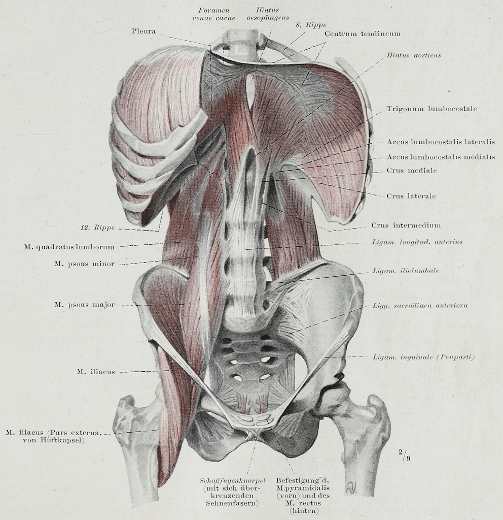
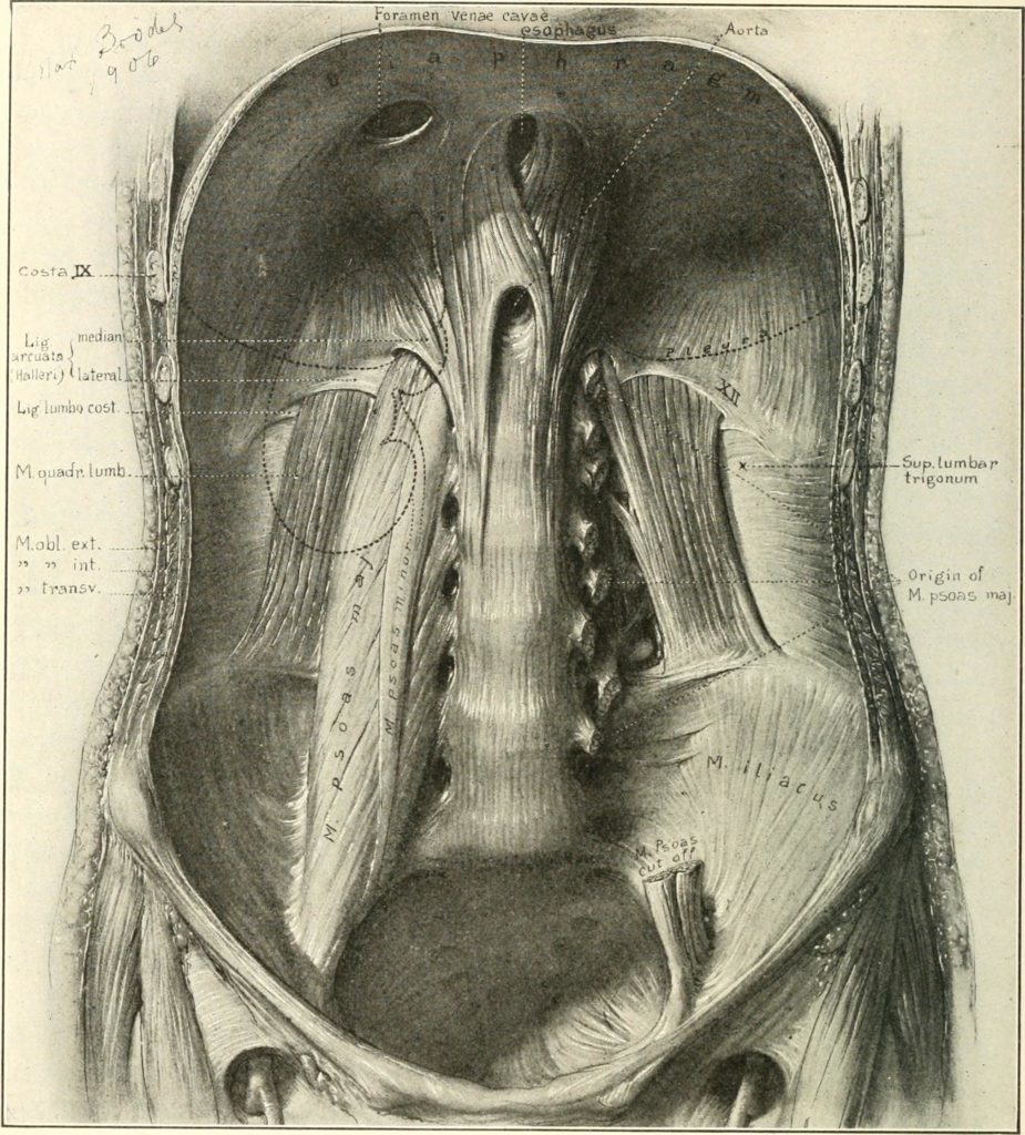
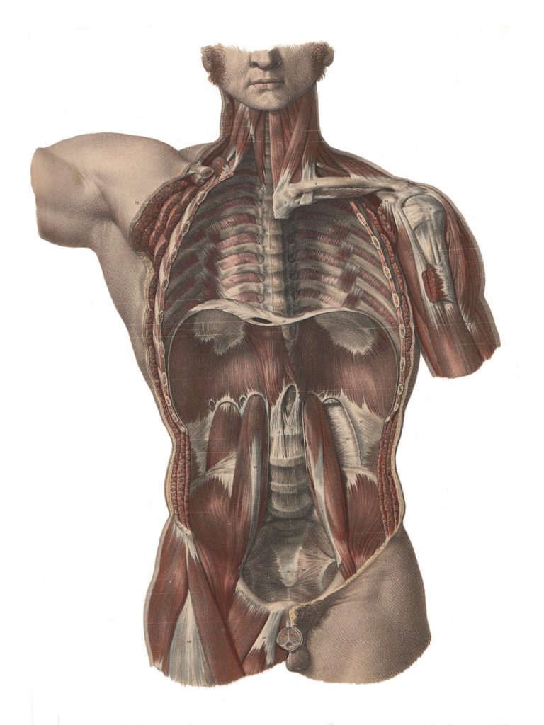
Anterior view of the diaphragm in situ, with complimentary views of the quadratus lumborum, the psoas major and minor, and iliacus. From Hermann Braus’ 1921 Anatomie des Menschen.Another anterior view of the diaphragm in situ. I like that this illustration shows the position of the subject’s right kidney (on the left side of the image), tucked up under the diaphragm in the back of the abdominal cavity. Not shown is the subject’s left kidney. Think about how much the kidneys have to move when the diaphragm descends on the inhalation. Illustration by Max Brödel, from Howard A. Kelly’s 1922 Diseases of the kidneys, ureters and bladder, with special reference to the diseases of women.The Diaphragm in situ, this time with a view of the thoracic cavity above the diaphragm, in addition to the abdominal cavity below. Note the proximity of the psoas muscles (the vertically-oriented muscles on either side of the spine). This illustration also offers a nice perspective on the diaphragm in relation to the muscles of the pelvic floor. Illustration by Nicolas-Henri Jacob, from Jean Baptiste Marc Bourgery’s Traité complet de l’anatomie de l’homme, comprenant la médicine, published in sixteen volumes between 1831 and 1854.
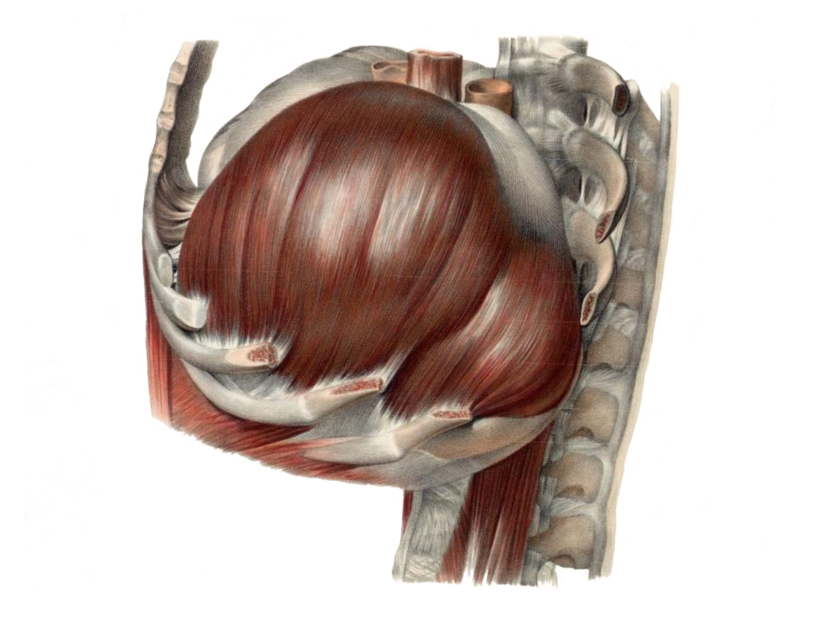
The Diaphragm Itself
It’s hard to beat Nicolas-Henri Jacob’s illustrations from Traité complet de l’anatomie de l’homme, Bourgery’s sixteen-volume work published between 1831 and 1854.
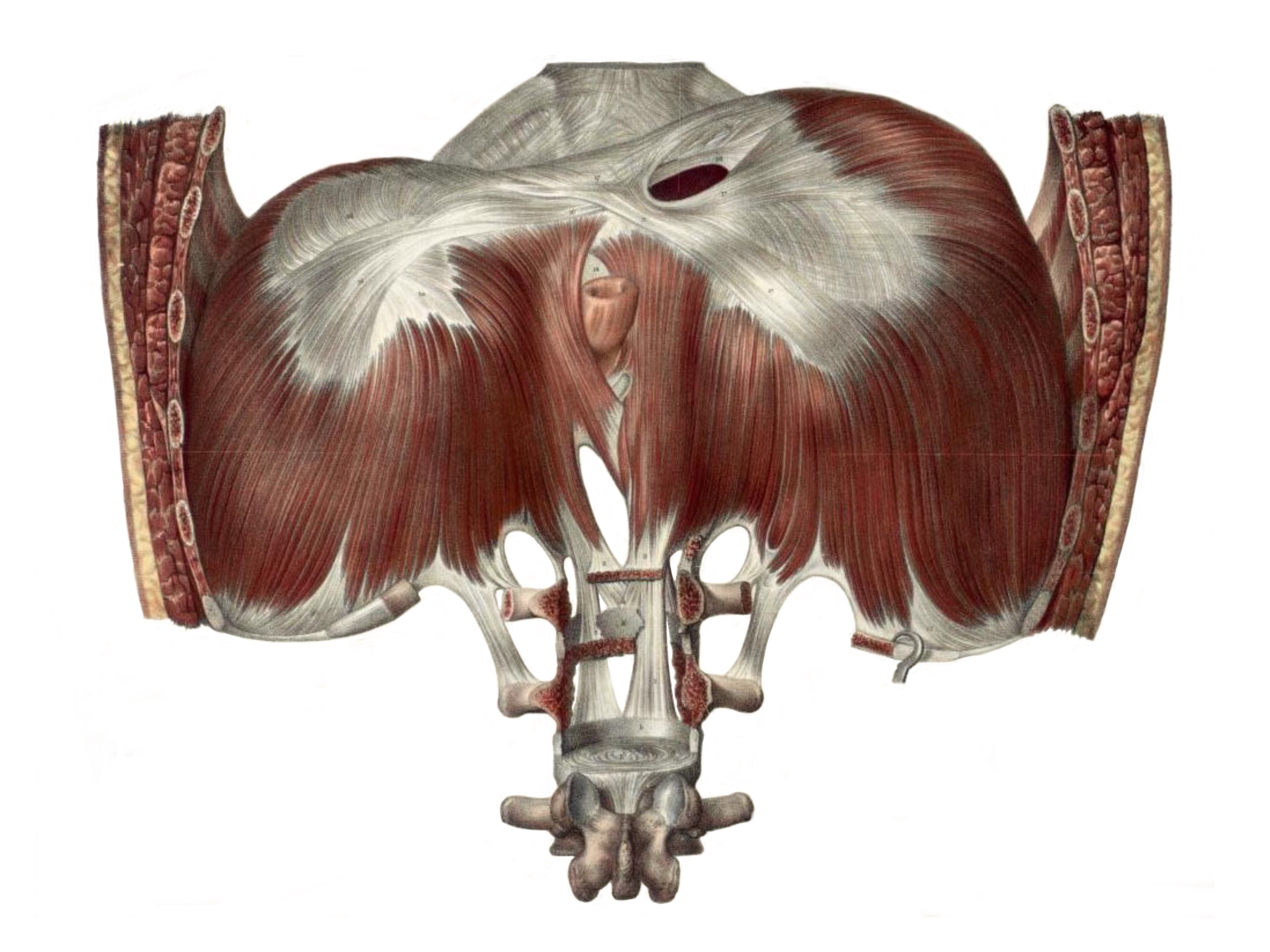
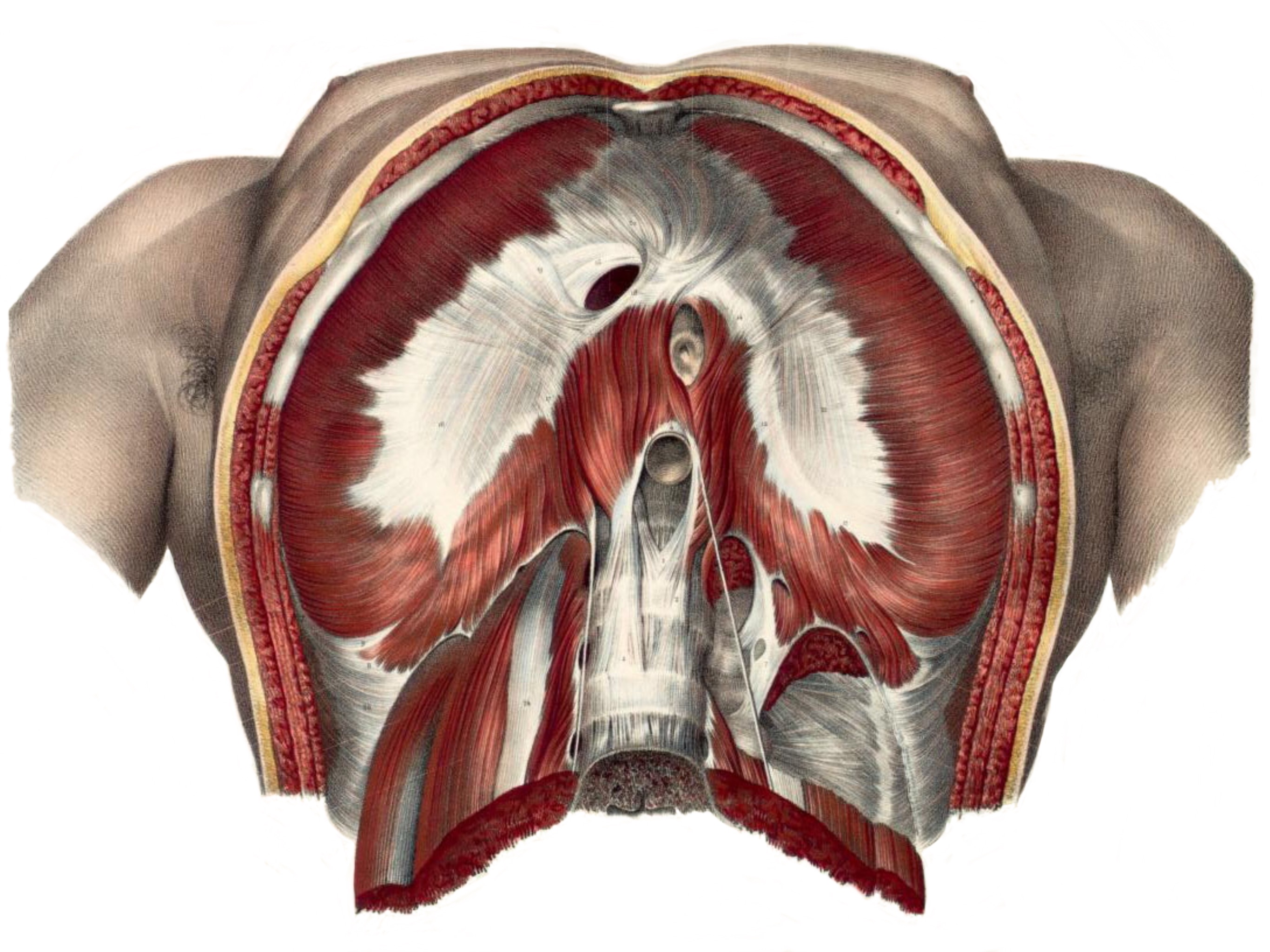
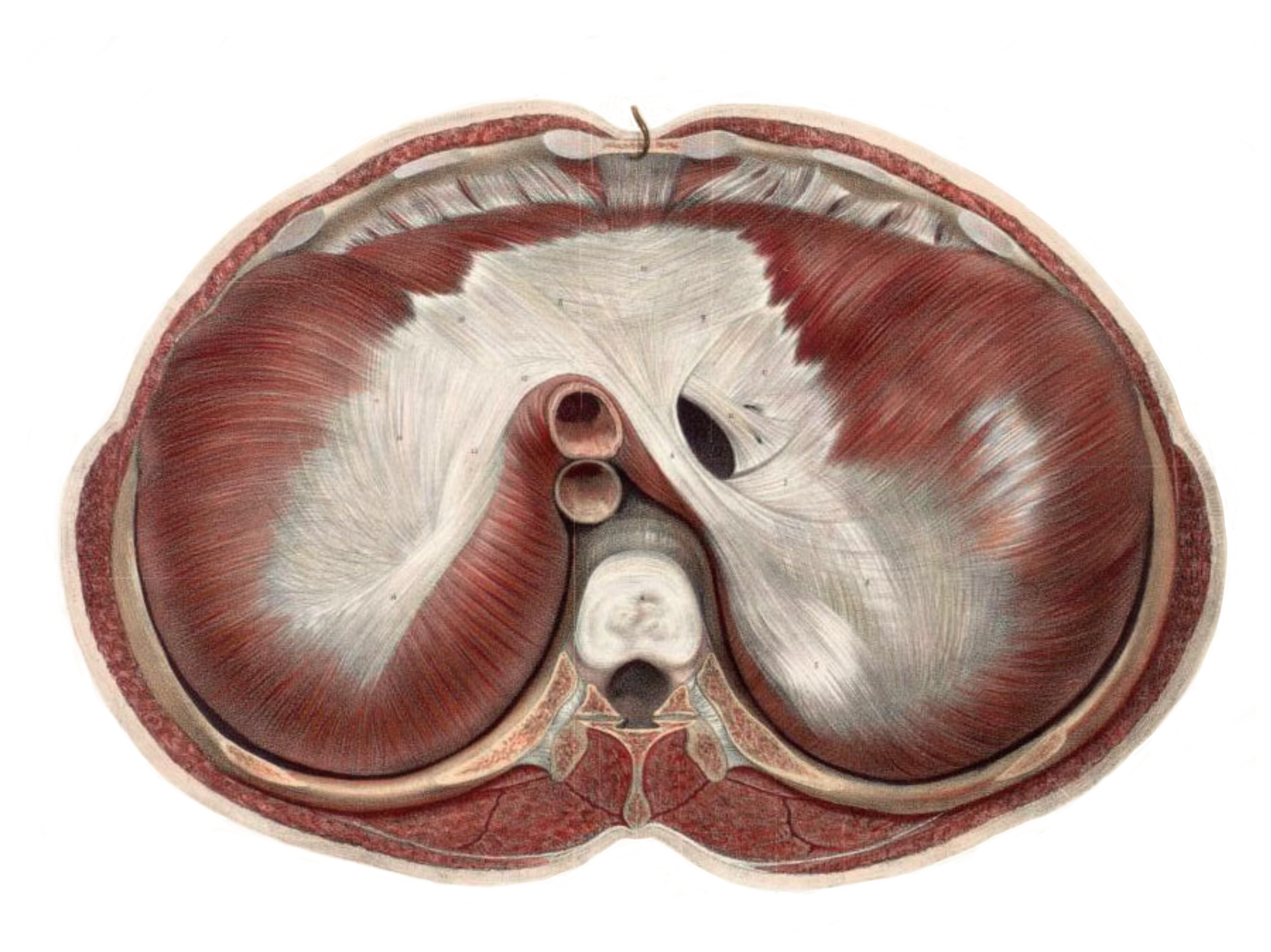
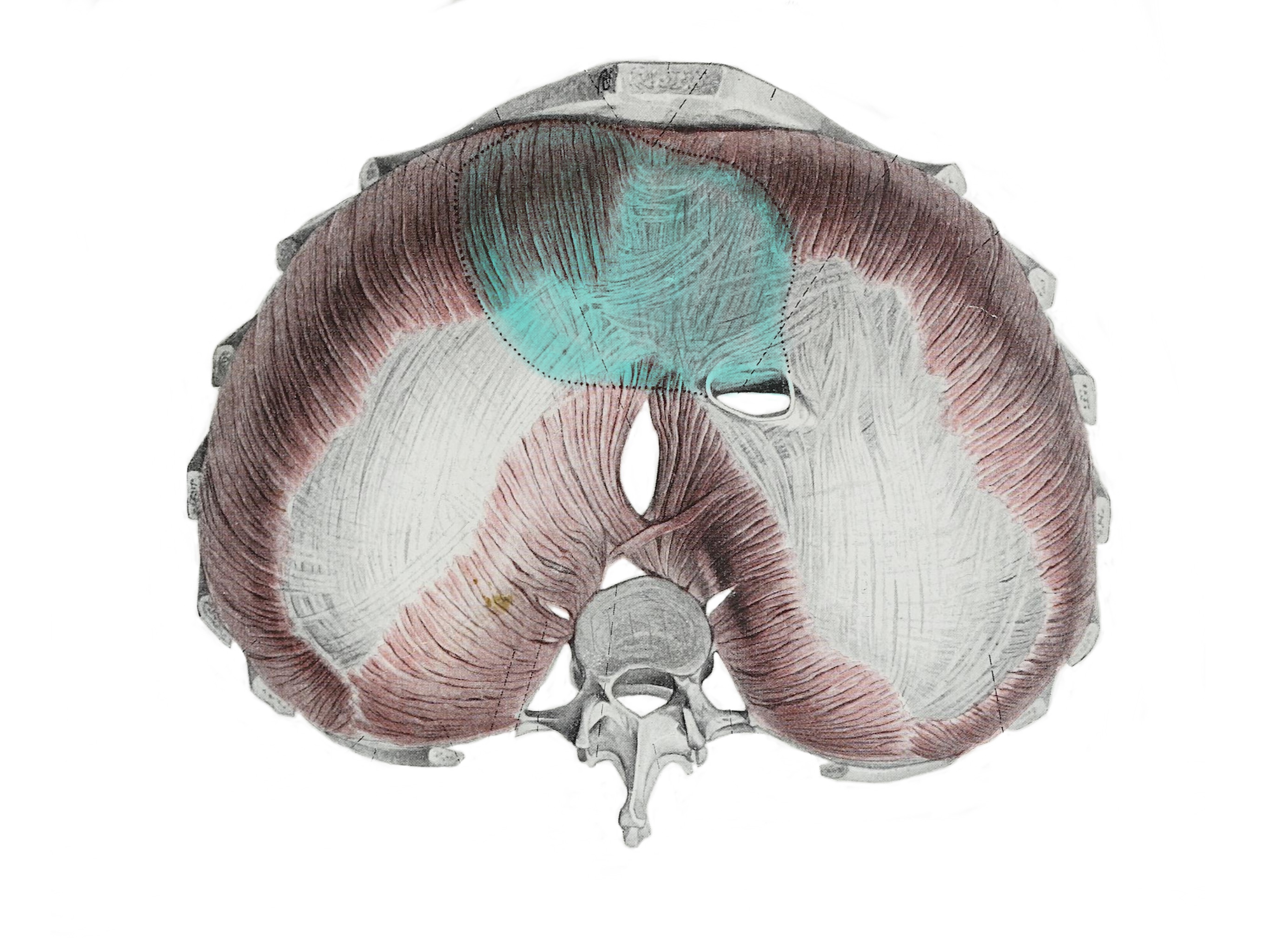
Anterior viewPosterior viewSagittal viewInferior viewSuperior view. Note, the front of the body is at the top of the image.Another superior view; the blue area shows where the pericardium (the bag around the heart) attaches to the top of the diaphragm. Illustration from Hermann Braus’ 1921 Anatomie des Menschen: ein Lehrbuch für Studierende and Ärzte.
What’s above and below the diaphragm?
The diaphragm is both the floor of the thoracic cavity, and the ceiling of the abdominal cavity. Since it’s constantly in motion — we breathe roughly twenty thousand times a day — it’s good to know what’s above and below it, so you can start to think about how the movement of the diaphragm effects everything around it.
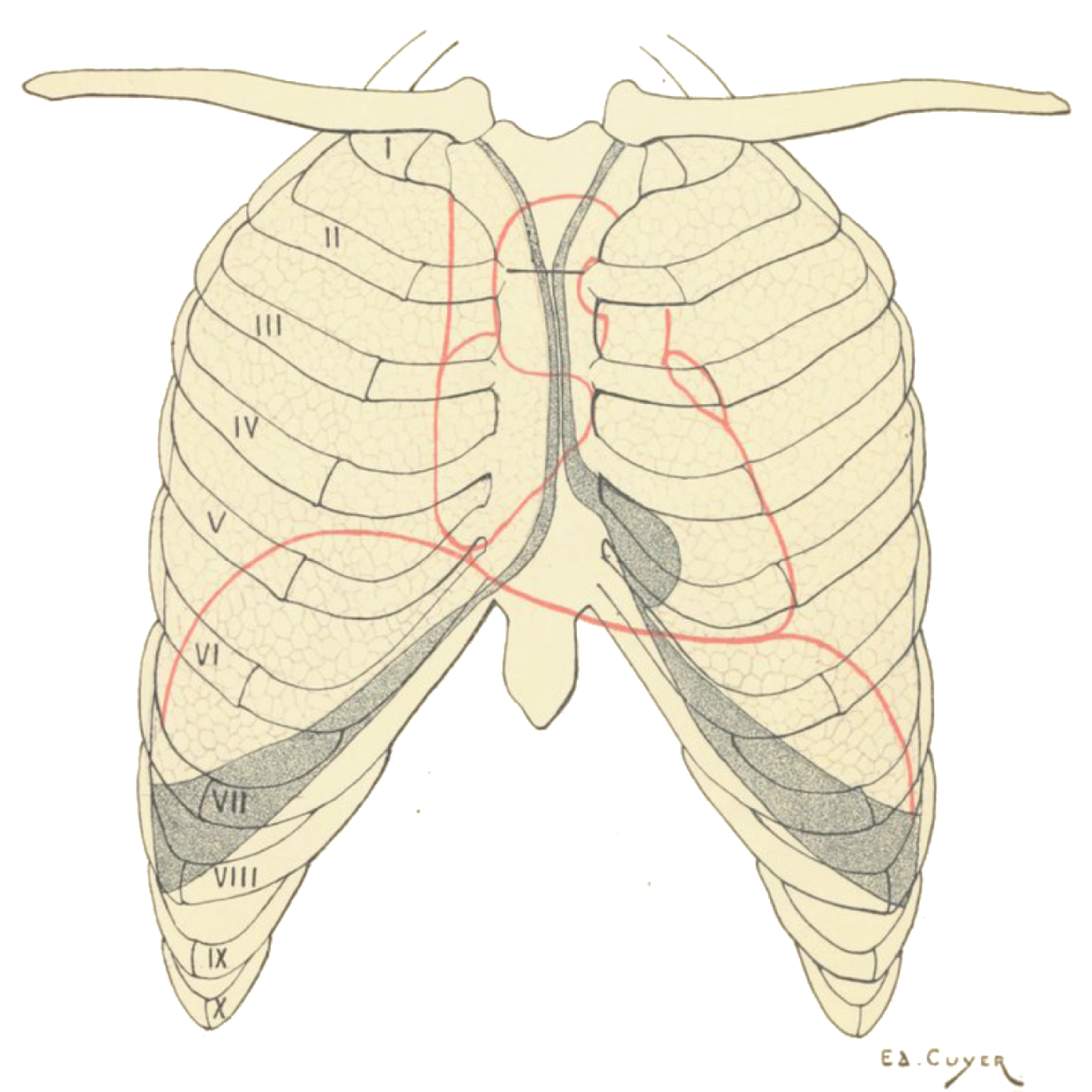
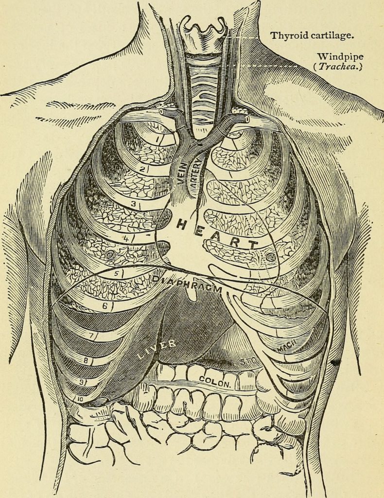
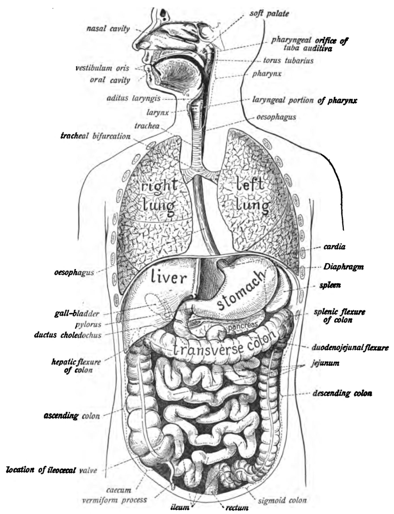
The diaphragm in situ with the abdominal organs. Note that the liver (on the subject’s right) and stomach are directly under the diaphragm, tucked up under the base of the ribcage. Below that is the transverse colon. Many singers, when asked to show where the top of their diaphragms are, will put their hands in the region of the transverse colon. The domes of the diaphragm are higher than you think.If you are inclined to count your ribs, you can get a sense of the level of the domes of the diaphragm, in the vicinity of the fifth and sixth ribs, a little higher on your right side. Illustration by Édouard Cuyer from Paul Poirier’s 1899 Traité d’anatomie humaine.Another useful view of the position of the diaphragm relative to the ribs and the upper abdominal organs. Illustration from the book with the longest title in the world: Frederick Garbit’s 1880 The woman’s medical companion and guide to health: a practical treatise on the diseases of women and children: with full and definite directions for their treatment, giving the causes, symptoms, and means of prevention or cure, with the latest and most approved methods of treatment adopted by all schools of medicine: their doses and modes of administration carefully prescribed.This guy has no heart (among other issues). But I like that he has a vocal tract, and that you can perhaps imagine how, when the diaphragm descends on the inhale, it pulls on the lungs, which pull on the trachea, which pulls on the larynx. Illustration from Johannes Sobatta’s 1906 Atlas of Human Anatomy.Beautiful illustration of the diaphragm and thorax by Patrick Lynch, generated for multimedia teaching projects by the Yale University School of Medicine, Center for Advanced Instructional Media, 1987-2000. I love that this illustration gives you a sense of the depth, front to back, at the base of the lung and top of the diaphragm. Shared under a Creative Commons Atribution 2.5 License.
The Diaphragm as it Moves
Most of the illustrations above are over 100 years old, and are necessarily static. More recent technology enables us to see images of the diaphragm and surrounding structures in movement, such as this real-time MRI of the movement of the thorax during breathing, made by the good folks at Biomedizinische NMR Forschungs GmbH.