Ah, January!
Seattle poet Greta Nintzel sometimes calls the skull a “calcium cage.” Until recently, I admired the phrase as an apt description for my own noggin’: a bony container for the wizard running my body machine.
Enter the craniosacral therapist. She put her hands on my head and made the subtlest of movements. I started to think: maybe I’m not as hard-headed as I thought.
Craniosacral therapy is based (in part) on the idea that the bones of the skull retain some amount of movement at the sutures where they grow together during development1. I have a picture in my mind of the half-baked, developing skull as a group of islands, slowly expanding towards one another until they can start to merge together, first as continents, and then as a whole, head-shaped planet.
Before the craniosacral therapist put her hands on my skull, I had no thought of there being movement in the skull itself, my oh-so-solid calcium cage. And now? I think of the bones as having slightly lubricated edges, as if they are moored to one another on the sea of my gray matter, bobbing and floating (while I dreamily listen to the halyards slapping against the mast).
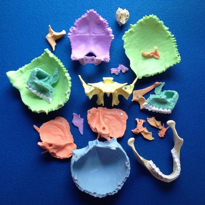
There is some disagreement on what should or shouldn’t be counted in this bony island archipelago, but for our purposes, let’s say there are 22 bones of the skull, as in this disarticulated model (Legos for anatomy geeks):
And here it is again, put back together:
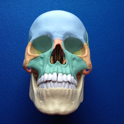
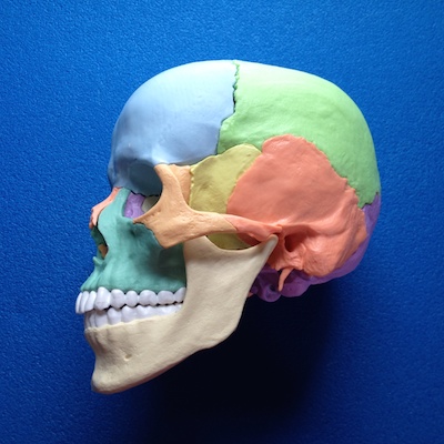
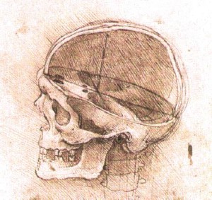
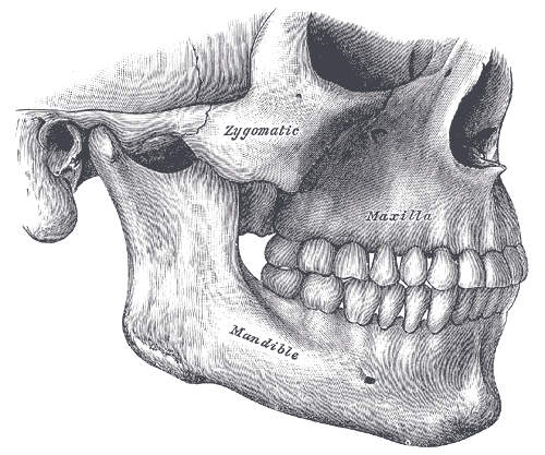
Among all these, I want to draw your attention to the maxillary bones, which are just below the nasal opening (teal in the images above). They hold your upper teeth and provide most of the surface of your hard palate. Here is another image from the 1918 edition of Gray’s Anatomy:
Previously, I introduced you to Joseph Stemple’s Vocal Function Exercises, and promised some things to contemplate while practicing them. So let’s vibrate the maxilla.
- As you hum, see if you can sense vibration in this bone (try different pitches). It’s easy to palpate its location, through the skin and muscles of your face, and also inside your mouth. If you feel the midline of your hard palate with your tongue, you can probably find the seam where the two halves of the maxilla join together. Can you feel the hum vibrating in different areas of the bone and in your upper teeth?
- The roof of your mouth is also the floor of your nose. Inhale slowly through your nose. Can you use the sensation of air coming in to help you sense the top (superior aspect) of the maxilla? What happens if you think of the hard palate widening slightly with the inhale?
- In the Vocal Function Exercises, we learned to sing long-tones on what I call the [o] inside an [u] vowel. Here, once again, is an example of the vowel:
Because the embouchure of the closed [u] vowel is so tight, it can be tiring to hold the position for the duration of the exercise. To counter this, I like to imagine I can relax my upper teeth, as if I’m “letting them go”, and let them buzz in their sockets a little.
And hey, once you’ve vibrated the maxilla, we’ve only 20 bones to go in our vibratory exploration of the bones of the skull (the maxilla counts as two bones, with left and right halves).
Have fun!